Cytokinesis, the final stage of cell division, is the process by which a single cell divides its cytoplasm to form two daughter cells. While the goal of cytokinesis is the same in both animal and plant cells—to ensure each daughter cell receives the necessary organelles and cytoplasmic components—the mechanisms differ significantly due to the structural differences between these cell types. Let’s dive into the fascinating world of cytokinesis and explore how it occurs in animal and plant cells.
Cytokinesis in Animal Cells
In animal cells, cytokinesis is characterized by the formation of a cleavage furrow. Here’s how it works:
- Formation of the Contractile Ring
During late anaphase or telophase, a ring composed of actin and myosin filaments forms just beneath the plasma membrane at the cell’s equator. These proteins are the same ones responsible for muscle contraction, and they play a key role in cytokinesis. - Contraction of the Ring
The actin-myosin ring contracts, pulling the plasma membrane inward. This creates a groove or indentation called the cleavage furrow, which gradually deepens as the ring tightens. - Pinching the Cell in Two
The cleavage furrow continues to contract until the cell is pinched into two separate daughter cells, each enclosed by its own plasma membrane. This process is often compared to tightening a drawstring around the waist of a bag. - Completion of Division
Once the division is complete, the two daughter cells may remain connected temporarily by a midbody structure, which eventually disassembles.
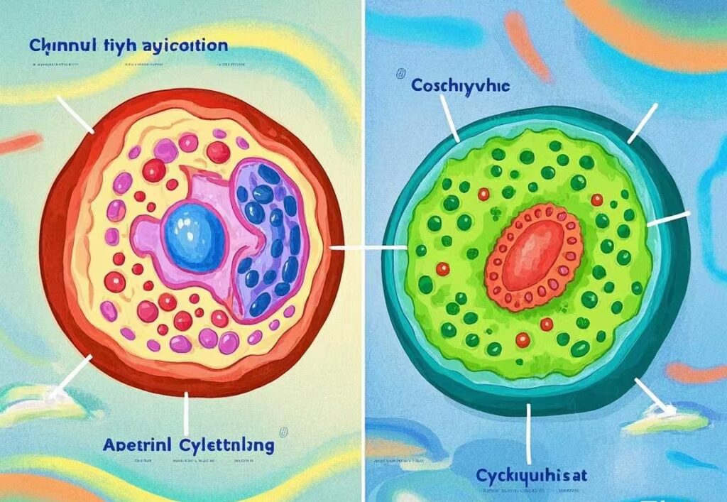
Cytokinesis in Plant Cells
Plant cells, on the other hand, face a unique challenge: they are surrounded by a rigid cell wall that prevents them from forming a cleavage furrow. Instead, plant cells use a different mechanism involving the formation of a cell plate. Here’s how it works:
- Formation of the Cell Plate
During telophase, vesicles derived from the Golgi apparatus and endosomes gather at the center of the dividing cell. These vesicles contain materials needed to build the new cell wall and plasma membrane. - Fusion of Vesicles
The vesicles fuse together at the equator of the cell, forming a disc-like structure called the cell plate. The cell plate grows outward, guided by microtubules of the phragmoplast (a structure unique to plant cells). - Development of the New Cell Wall
As the cell plate expands, it merges with the existing plasma membrane and cell wall, effectively dividing the cell into two. The cell plate eventually matures into a new cell wall, separating the two daughter cells. - Completion of Division
Once the cell plate is fully formed, the two daughter cells are completely separated, each enclosed by its own cell wall and plasma membrane.
Key Differences Between Animal and Plant Cytokinesis
| Aspect | Animal Cells | Plant Cells |
|---|---|---|
| Mechanism | Cleavage furrow formed by actin-myosin ring | Cell plate formed by vesicle fusion |
| Structure Involved | Contractile ring | Phragmoplast and Golgi-derived vesicles |
| Cell Wall | Absent; flexible plasma membrane | Present; rigid cell wall prevents pinching |
| Energy Requirement | High (due to actin-myosin contraction) | Moderate (vesicle transport and fusion) |
| Final Outcome | Two daughter cells with identical membranes | Two daughter cells with new cell walls |
Why the Difference?

The differences in cytokinesis between animal and plant cells are largely due to their structural and functional adaptations:
- Animal cells rely on flexibility and mobility, which is why they use a contractile ring to pinch the cell in two.
- Plant cells require rigidity and support, provided by their cell walls. The cell plate mechanism allows them to build a new wall while dividing.
Conclusion
Cytokinesis is a remarkable process that highlights the diversity of life at the cellular level. While animal and plant cells achieve the same goal—successful cell division—they do so in ways that reflect their unique biological needs. Whether it’s the contracting cleavage furrow of an animal cell or the growing cell plate of a plant cell, cytokinesis is a testament to the elegance and complexity of cellular biology.